


Patient Resources
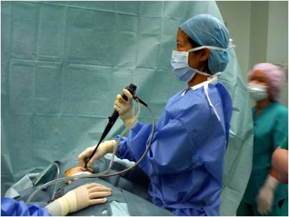
Medical Thoracoscopy / Pleuroscopy
What is Pleuroscopy?
A pleuroscopy is a medical procedure that allows the doctor to visualise the pleural cavity, which is the space between the chest wall and the lung. This is done by inserting a thin tube with a camera, called a pleuroscope, through the chest wall.
When is a Pleuroscopy usually performed?
Pleuroscopy is usually performed to examine and diagnose the cause of abnormal fluid accumulation in the pleural cavity. It allows visualisation of the pleural surface and enables biopsies under direct vision. During the procedure, the doctor may occasionally spray medications onto the pleural surface to prevent the fluid from re-accumulating.
How is a Pleuroscopy performed?
A pleuroscopy is usually performed under conscious sedation in the endoscopy suite or in the operating theatre. This means that the patient will be given a medication to make them sleepy and to help them relax. Occasionally, the doctor may decide to perform this procedure under general anaesthesia.The patient will be asked to lie on the side. The area where the pleuroscope will be inserted is sterilised with cleaning solutions. A pleuroscope is then inserted then into the chest. An ultrasound is sometimes used to locate the most appropriate port of entry. Any fluid seen is drained out and the pleural cavity is then carefully inspected. Biopsies will be performed if there are abnormal areas in the pleural space.Towards the end of the procedure, medications may be sprayed into the pleural space to prevent re-accumulation of fluid. A drainage tube may be left behind, to allow any fluid or air to drain out over the next few days.
What are the complications of a Pleuroscopy?
Complications of medical thoracoscopy/pleuroscopy are uncommon. They may include bleeding, infection of the pleural space or injury to the internal organs of the chest. Occasionally, the lungs may fail to expand properly due to prolonged drainage of air or fluid.
Preparing for Pleuroscopy
Before performing a pleuroscopy, the doctor will talk to the patient about the procedure, the risks involved and what he/she may expect to find. The patient will be asked to sign a consent form. If the patient is on any blood-thinning medications, such as aspirin, clopidogrel (Plavix) or warfarin (Coumadin), he/she may be asked to discontinue these for a period of time before the test. In addition, the patient will be asked to fast (not eating or drinking) for 4-6 hours before the procedure.
What happens after the procedure?
Following the procedure, the patient will have a chest drain to drain out both air and fluid. This is to facilitate lung re-expansion. Pain medication will be routinely given to help manage pain. When the lung has expanded and the drainage of air or fluid is minimal, the tube will be removed and the incision closed with a stitch. After discharge, the patient will be asked to come back in approximately one week for review and to have the stitch removed.